When a patient comes into my office complaining of heel pain, one of the first things we discuss is whether a heel X-ray is necessary. I often hear, "Doctor, I have this pain that won't go away, especially in the morning."
A heel X-ray—or heel x-ray, as we often say—isn't always the first thing to do. But when is it needed? And above all, what can it tell us that's useful? Let's explore these aspects together, because understanding when it's truly necessary can make a difference in your treatment journey.
The calcaneus: much more than just a heel bone
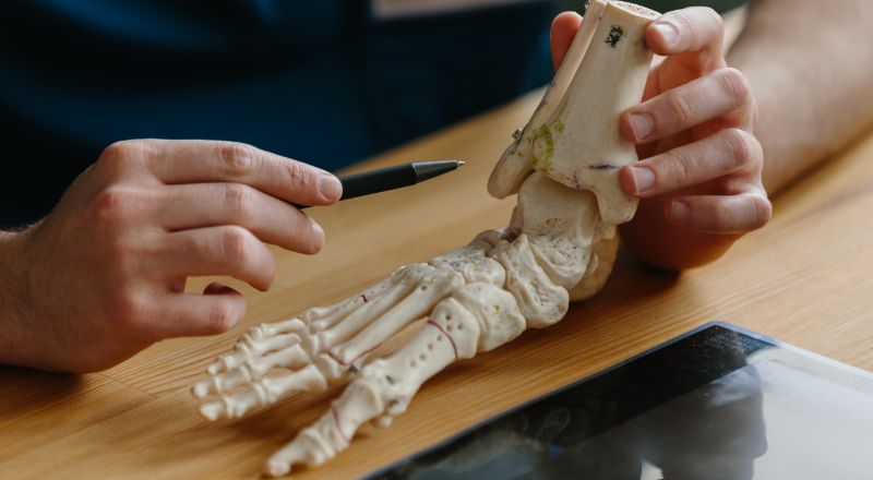
The calcaneus is the largest bone in the foot. Think about it: it has to support your entire body weight with every step. It has a unique shape, with multiple articular surfaces that connect with the talus above and the cuboid in front.
Its anatomy is complex. The posterior tubercle, where the Achilles tendon attaches, the facet joints that allow for foot movement, and the plantar tuberosity, where the plantar fascia begins. Even the slightest alteration in these areas can cause bothersome symptoms.
This is why it's sometimes crucial to "see inside" with a heel X-ray. The problem isn't always obvious from the outside.
When I suggest you take a heel x-ray

I never do an X-ray just for the sake of it. There are specific indications that push me to request one, and now I'll explain them to you just as I explain them to my patients.
Fractures are obviously the first thing we think of after a trauma. The other day, a patient came to me who had fallen from a ladder—in these cases, a heel X-ray is mandatory to rule out bone lesions.
But there are also less dramatic situations that require further investigation. Heel spurs, for example. It's that small bony growth that forms where the plantar fascia attaches and can cause severe pain, especially in the morning. Only a heel x-ray can clearly see it.
Then we have osteoarthritis of the calcaneal joints, congenital bone deformities, and in rarer cases, we must rule out bone infections or tumors. In all these situations, the heel X-ray becomes a fundamental diagnostic tool.
How the exam is conducted
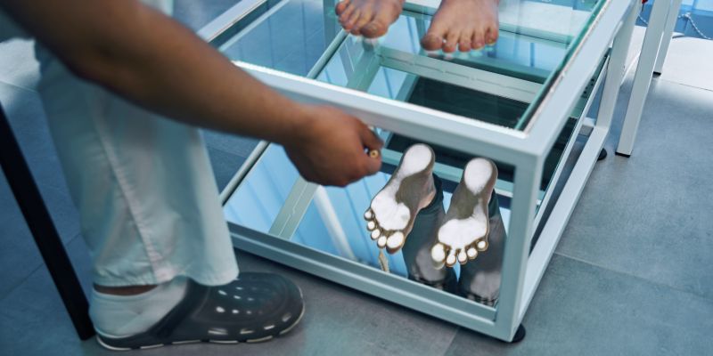
I always tell my patients, “There's nothing to worry about.” The procedure is quick and easy.
You position yourself on the x-ray table, and the technician asks you to hold your foot in specific positions—usually one lateral and one downward. It lasts just a few minutes. There's no pain, just the discomfort of having to hold the position for a few seconds.
No special preparation is needed. Just remove your shoes and any socks, and you're done. The only thing you'll feel is perhaps a little pressure if you have to rest your sore foot on the support, but only for a few moments.
How do I interpret the results?
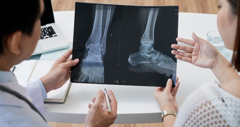
When I receive your heel X-ray images, I look at several elements. First, I check the bone integrity—are there any fractures? Then I examine the joint margins for signs of arthritis.
An important aspect is comparison with any previous X-rays. The evolution of a disease over time tells us a lot about how you're responding to treatment and whether we need to change our approach.
I look for signs of abnormal calcifications, such as the famous heel spur, changes in bone density, or thickening of the surrounding soft tissue. Every little detail has its diagnostic significance.
To be honest, I always interpret the images in conjunction with your medical history and the physical examination. An X-ray alone, without context, can tell us little or even be misleading.
The limits we need to know
Heel X-rays have limitations, and you should be aware of them. They show bones well, but soft tissue—tendons, ligaments, fascia—is virtually invisible.
If I suspect an Achilles tendon injury or acute plantar fasciitis, I often prefer an ultrasound or MRI. I use a CT scan when I need more precise bone details, perhaps before surgery.
In short, a heel x-ray is an excellent starting point, but it's not always the definitive test. Each situation requires the most appropriate diagnostic approach.
Safety and precautions
The radiation used is minimal, truly negligible. To give you an idea, it's like spending a few extra hours in the sun during the summer.
The only special care we take is for pregnant women. If you are pregnant or suspect you are, always let me know before the exam. In these cases, we consider alternatives such as ultrasound, which does not use ionizing radiation.
The SmartHallux approach for heel pathologies

Here at SmartHallux, we've developed specific minimally invasive techniques to treat many heel pathologies that we often discover through x-rays. Dr. Luigi Manzi has gained significant experience in this field.
Whether you're dealing with chronic plantar fasciitis, symptomatic heel spurs, or Achilles tendon problems, our techniques allow for faster and less painful recoveries than traditional approaches.
The beauty of minimally invasive surgery is that we can often resolve seemingly complex problems with small, targeted surgical procedures. Of course, this always follows a careful evaluation, starting with a good heel X-ray.
We primarily treat conditions such as hallux valgus , Morton's neuroma , and other deformities that cause foot pain. Providing comprehensive support throughout the treatment process, SmartHallux can be a winning choice for effective treatment tailored to your needs. Book your specialist consultation with the SmartHallux team now.
FAQ
Does the X-ray hurt?
Not at all. The only discomfort might be holding the position if you're in a lot of pain, but we're talking seconds.
How much does a heel x-ray cost?
With the national health system, it's free with a referral. Privately, the costs are affordable, and it's often a worthwhile investment for quick answers.
How long will it take to see the results?
You usually get the report within 24-48 hours. Except for emergencies, where the waiting time is reduced to a few hours.
Do I need to do anything before the exam?
Nothing special. Just bring any previous X-rays for comparison.